
Optical Coherence Tomography (OCT) – Eye Imaging Test
 Optical Coherence Tomography (OCT) is a new type of imaging technology. OCT is used for taking cross-sectional pictures of the retina. The retina is located at the back of your eye. It is used to diagnose and follow treatment in certain eye conditions and diseases, such as age-related macular degeneration, diabetic retinopathy and other diseases affecting the macula.
Uses
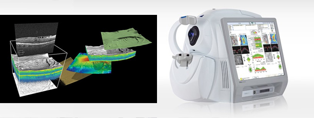
OCT is a non-invasive imaging test. OCT is a unique test because it allows doctors to look at cross-sectional layers of the eye. OCT can be used to screen for, diagnose, and monitor conditions of the retina and optic nerve.
Each of the ten layers in the retina can be detected. OCT allows a doctor to measure the thickness of each layer to aid in the early detection and diagnosis of retinal diseases and conditions. Such conditions include age-related macular degeneration, diabetic retinopathy, and many others.
Detailed information can also be obtained about he optic nerve. OCT allows a doctor to measure the thickness and configuration of the optic nerve to aid in the early detection and diagnosis of optic nerve disorders. Such conditions include glaucoma, optic neuritis, papilledema (optic nerve swelling that may be caused by things such as brain tumors or other conditions) and many others.
Preparation
There is no special preparation for this test.
Testing
The testing time is very short. You will be seated for the test. Your face will be stabilized with a positioning device during the test. The technician will use a special camera to take pictures of your inner eye. The images are transmitted to a computer for Dr. Goff to view.