Available Cutting Edge Diagnostic and Advanced Screening Services
River Country Eye Care offers the following diagnostic technology:
-
Digital Retinal Photography and OptoMap Retinal Imaging
-
External Ocular Photography
-
Optical Coherence Tomography (OCT)
-
Automated Threshold Visual Field Testing
-
Pachymetry
-
Gonioscopy
-
RPS Adeno Detector for Diagnosing Adenoviral Conjunctivitis
-
TearLab Tear Osmolarity Testing for Dry Eye Disease
Optomap ultra-wide digital retinal imaging


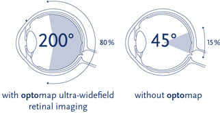
The optomap ultra-widefield retinal image is a unique technology that captures more than 80% of your retina in a single image while traditional imaging methods typically only show 15% of your retina at one time. The optomap ultra-wide digital retinal imaging system helps you and your eye doctor make informed decisions about your eye health and overall well-being.
Your retina (located in the back of your eye) is the only place in the body where blood vessels can be seen directly. This means that in addition to eye conditions, signs of other diseases (for example, stroke, heart disease, hypertension and diabetes) can also be seen in the retina. Early signs of these conditions can show on your retina long before you notice any changes to your vision or feel pain. While eye exams generally include a look at the front of the eye to evaluate health and prescription changes, a thorough screening of the retina is critical to verify that your eye is healthy.
Benefits of an optomap
The benefits of having an optomap ultra-widefield retinal image taken are:
- optomap facilitates early protection from vision impairment or blindness
- Early detection of life-threatening diseases like cancer, stroke, and cardiovascular disease
- The unique optomap ultra-widefield view helps Dr. Goff detect early signs of retinal disease more effectively and efficiently than with traditional eye exams.
- Unlike traditional retinal exams, the optomap image can be saved for future comparisons.
Early detection means successful treatments can be administered and reduces the risk to your sight and health.
Preparation
There is no special preparation for this test. You may have your pupils dilated for this test but many times this is not necessary.
Testing
Getting an optomap image is fast, painless and comfortable. Nothing touches your eye at any time. It is suitable for the whole family. To have the exam, you simply look into the device one eye at a time (like looking through a keyhole) and you will see a comfortable flash of light to let you know the image of your retina has been taken.
Under normal circumstances, dilation drops might not be necessary, but Dr. Goff will decide if your pupils need to be dilated depending on the health of your eyes. The image capture takes less than a half second and they are available immediately for you to see your own retina. You see exactly what Dr. Goff sees – even in a 3D animation.
Optical Coherence Tomography (OCT) – Eye Imaging Test


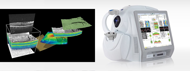
Optical Coherence Tomography (OCT) is a new type of imaging technology. OCT is used for taking cross-sectional pictures of the retina (located in the back of your eye). It is used to diagnose and follow treatment in certain eye conditions and diseases, such as age-related macular degeneration, diabetic retinopathy and other diseases affecting the macula.
Benefits of an OCT
OCT is a non-invasive imaging test. OCT is a unique test because it allows doctors to look at cross-sectional layers of the eye. OCT can be used to screen for, diagnose, and monitor conditions of the retina and optic nerve.
Each of the ten layers in the retina can be detected. OCT allows a doctor to measure the thickness of each layer to aid in the early detection and diagnosis of retinal diseases and conditions. Such conditions include age-related macular degeneration, diabetic retinopathy, and many others.
Detailed information can also be obtained about he optic nerve. OCT allows a doctor to measure the thickness and configuration of the optic nerve to aid in the early detection and diagnosis of optic nerve disorders. Such conditions include glaucoma, optic neuritis, papilledema (optic nerve swelling that may be caused by things such as brain tumors or other conditions) and many others.
Preparation
There is no special preparation for this test. You may have your pupils dilated for this test but many times this is not necessary.
Testing
The testing time is very short. You will be seated for the test. Your face will be stabilized with a positioning device during the test. The technician will use a special camera to take pictures of your inner eye. The images are transmitted to a computer for your doctor to view.
